Traumatic brain injury (TBI) can occur when the head is hit with a sudden, blunt force—think a car accident or sports injury.
While many symptoms of TBI are obvious, others are less so. For example, one may not immediately notice that the size and shape of their pupil have changed after experiencing a TBI.
Therefore, it is important for doctors to be familiar with the pupillary response in traumatic brain injuryand all its implications.
Pupilometry
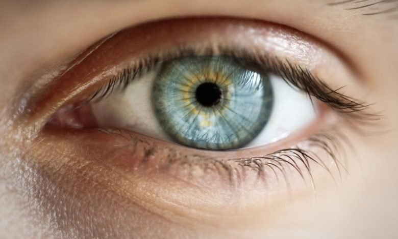
Pupilometry is a medical test that measures the size and reaction of your pupils.
When you go to see an eye doctor, they will use a pupilometer to measure pupil dilation. This can help diagnose neurological conditions like brain injury, stroke, and seizures. Pupils are the black circles inside your eyes that allow light into them so they can see. Your pupils react to changes in light by constricting or dilating in response to how bright or dark it is around them.
This lets you see objects in different lighting conditions without having to adjust the amount of light entering your eyes (which would be difficult).
Pupillary Light Reflex Testing
Doctors can easily assess a patient’s pupillary light reflex by shining a small, bright light into one eye.
The pupil that is being illuminated should contract in response to the light source. It is important to record the amount of time it takes for this response to occur. If it takes too long, the patient may have an issue with their pupil’s ability to react appropriately to changes in lighting conditions and ambient lighting levels.
Types of Pupil Reaction
- Constriction: The pupil constricts or closes when exposed to light. This is called an afferent response because it occurs when the pupil is stimulated.
- Miosis: The opposite of constriction, miosis is a reflexive response that makes the pupils dilate (open) when exposed to light or other stimulation.
- Mydriasis: Experts also refer to this as “dilatation of the pupil,” and it happens when you have an injury or head trauma, such as a concussion or brain injury. It can happen right away but often takes several hours after the doctor can notice it.
- Anisocoria: When one pupil does not normally react—like during a concussion—you may notice that one eye seems larger than the other; this means there was some damage to their vision and/or brain function in that particular area of their body.
Constriction velocity
Pupil reaction time is the time it takes for the pupil to constrict in response to light.
It’s measured in seconds and is used as a sign of how quickly information is being processed by your brain. The normal pupil reaction time is 1-2 seconds. For example, if a doctor shines a flashlight at a patient’s eye and then measures how long it takes for their pupil to shrink again, that would be their normal baseline (though this measurement can vary slightly from person to person).
Age, drugs and head injury affect pupil reaction times—but there’s no need for alarm if the patient falls outside of those ranges!
Understand the basics of pupil reactivity after head injury.
When a patient is in a dark room. They can still see their surroundings because of the light coming from outside.
The same is true for how we see with our eyes. There must be some light for us to be able to see. When a person’s pupils are dilate, it means that they have been expose to light. The opposite is also true—if their pupils are constrict or “contract,” then they haven’t been expose to enough light.
This process works like this: when more photons (particles of visible light) enter through the retina at one time than normal, the rods and cones release chemicals called neurotransmitters into nerve fibers leading from these cells toward an area called the thalamus in order for them to relay information about what type of stimulus has caused this reaction.
Meanwhile, ipsilateral parasympathetic nerves send signals back down towards. The lateral geniculate nucleus (LGN), located between the optic chiasm. Where retinal ganglion cell axons meet lateral geniculate nucleus axons before synapsing onto neurons in the anterior pretectal area behind the superior colliculi.
These projects toward the hypothalamus and brain stem centers, controlling pupil size via the parasympathetic system.
So, as long as someone doesn’t suffer physical damage during head trauma. Then they should have normal reactivity patterns upon re-exposure during the recovery period, regardless of whether they’re conscious or unconscious.